The microscope Levenhuk MED 30 is a trinocular microscope belonging to the professional series Levenhuk MED. The microscope uses infinity-corrected semi-flat achromatic optics, which allow the installation of additional accessories (available separately) to extend the performance of the microscope. This microscope is excellent for a specialist working in a clinical and diagnostic laboratory, research institute or university department. This microscope allows working with small structures and performing complex microbiological investigations
The microscope Levenhuk MED 30 is a trinocular microscope belonging to the professional series Levenhuk MED. The microscope uses infinity-corrected semi-flat achromatic optics, which allow the installation of additional accessories (available separately) to extend the performance of the microscope. This microscope is excellent for a specialist working in a clinical and diagnostic laboratory, research institute or university department. This microscope allows working with small structures and performing complex microbiological investigations
The microscopes of the Levenhuk MED 30 series are equipped with an infinity-corrected optical system that is typical for professional and high quality microscopes. This system includes infinity-corrected semi-flat objectives and allows for clear, high-contrast images with a high level of flatness.
One of the most important features of an infinity-corrected optical system is that it allows additional components to be installed in the optical path between the objective lens and the eyepiece tube. These additional components include polarizers, epi-fluorescence light and DIC prisms that exhibit minimal focusing deviations and correct aberrations. In summary, their modular design and ease of use make the Levenhuk MED 30 microscopes ideal optical instruments for use in different types of microscopy and work in hematology, histology, microbiology and other laboratories.
A trinocular head consists of an eyepiece tube for mounting a digital camera (not included) and a binocular head for visual observations. The 30° tilt angle is practical for prolonged work and the 360° rotating head allows effective use of the microscope for group work.
Wide field eyepieces allow diopter adjustment to the user's vision. The eyepieces provide 10x magnification. The microscope includes five objectives, the most powerful of which are equipped with retractable housings to protect the optics against accidental damage. The 100x objective is used for oil immersion observations. All objectives are semi-planar achromatic, which improves color reproduction, eliminates optical aberrations and provides a field of view with a high level of flatness. The accessory optics are protected with an anti-fungus coating. Sharpness is adjusted with the coarse and fine focus adjustment knobs.
The stage is equipped with a mechanical micrometer. The micrometer facilitates the arrangement of the specimens under the objective. Below the stage is an Abbe condenser with an iris diaphragm and a filter holder. Three light filters are included to enhance the image contrast and allow discerning small details of the specimens observed at high magnification. A 3-watt LED lamp is used to illuminate the specimens. The lamp is located under a condenser and its brightness can be adjusted. The LED lamp has a lens that enhances the brightness. Köhler illumination can be configured. The illumination is powered by an AC power supply.
Features:
- Trinocular head, magnification range 40x to 1000x
- Semi-flat, infinity corrected achromatic optics
- Anti-fungus coated eyepieces and objectives
- Optimized 3 watt LED lamp with brightness adjustment
- Köhler illumination possible
- Powered by AC power supply
Kit includes:
- Microscope base and stand
- 360° rotatable trinocular head
- Semi-flat infinity corrected achromatic objectives: 4x, 10x, 40xs, 60xs, 100xs (oil immersion) with antifungal coating
- Wide field eyepieces: WF10x/22 mm with antifungus coating (2 pcs.)
- Abbe N.A. 1.25 condenser with iris diaphragm and filter holder
- Filters: blue, green, yellow
- Immersion oil tube
- 3 watt LED lamp
- Fuse (2 pcs.)
- Microscope power cord
- Protective cover
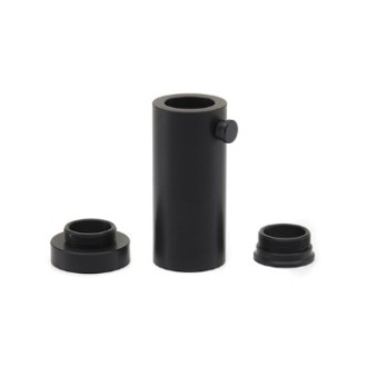
- Camera mount
- User manual and lifetime warranty
Caution:
Refer to the specification table for proper mains voltage and never attempt to plug a 110V device into a 220V outlet or vice versa without using a converter. Remember that the mains voltage is 110V in the United States and Canada, and 220-240V in most European countries.
Some of the things that can be seen under the microscope:


You can find these interesting slides and many more in the prepared slide sets at Levenhuk.
The binocular microscope Levenhuk MED 30T is compatible with digital cameras Levenhuk (available separately). The Levenhuk camera is installed in the eyepiece tube, replacing the eyepiece.
| Brand | Levenhukinc., USA |
| Warranty, years | lifetime |
| Package size (LxWxH), cm | 62x35x28 cm |
| Shipping weight, | 10.4 |
| Type | biological |
| Head | trinocular |
| Prism material | optical glass with antifungal coating |
| Nozzle | 360° rotatable |
| Head inclination angle | 30° |
| Magnification, x | 40-1000 |
| Eyepiece tube diameter, mm | 30 mm (binocular head), 23.2 mm (third vertical tube) |
| Eyepieces | WF10x/22 mm, wide field with diopter adjustment (2 pcs.) |
| Objectives | semi-flat achromatic infinity corrected objectives: 4x, 10x, 40xs, 60xs, 100xs (oil immersion) |
| Revolver | for 5 objectives |
| Interpupillary distance, mm | 48-75 |
| Stage, mm | 180x160 |
| Stage shift range, mm | 80/50 |
| Platen | mechanical, two-layer, with mechanical micrometer |
| Dioptric adjustment | ±5 |
| Condenser | Abbe N.A. 1.25 with iris diaphragm and filter holder |
| Diaphragm | iris |
| Focus | coaxial, coaxial, coarse (0.5 mm) and precise (0.002 mm), with rack-and-pinion system |
| Body material | metal |
| Illumination | LED |
| Adjustable brightness | True |
| Power supply | 100-240V |
| Type of light source | 3 watts, with an additional lens |
| Filters | green, blue, yellow |
| Additional | collector lens, Köhler illumination |
| Application | laboratory/medical |
| Location of illumination | bottom |
| Method of investigation | bright field |


