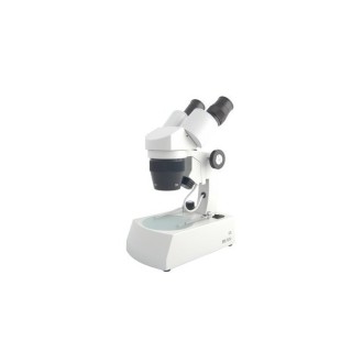
The trinocular microscope Levenhuk MED D45T is equipped with a 16 MP digital camera that allows taking pictures and recording high-resolution videos. The microscope is suitable for phase contrast observations as well as brightfield and darkfield observations. It is the perfect instrument for a clinical diagnostic center, biochemical laboratory or microbiological laboratory. The microscope allows the Köhler illumination to be configured; observations are made at a magnification of 40x to 1000x.
The trinocular microscope Levenhuk MED D45T is equipped with a 16 MP digital camera that allows taking pictures and recording high-resolution videos. The microscope is suitable for phase contrast observations as well as brightfield and darkfield observations. It is the perfect instrument for a clinical diagnostic center, biochemical laboratory or microbiological laboratory. The microscope allows the Köhler illumination to be configured; observations are made at a magnification of 40x to 1000x.
Microscopes of the series Levenhuk MED 45 microscopes are equipped with an infinity corrected optical system used in professional and high quality microscopes. This system includes infinity-corrected plano achromatic objectives that produce clear, high-contrast images with a high level of flatness.
One of the most important features of an infinity-corrected optical system is that it allows any additional elements to be installed in the path between the objective lens and the eyepiece lens. Additional elements include polarizers, epifluorescence light and DIC prisms that exhibit minimal focusing deviations and correct aberrations. In summary, their design and ease of use make the microscopes Levenhuk MED 45 optical instruments are ideal for use in different types of microscopy and work in hematology, histology, microbiology and other laboratories.
The trinocular head consists of the binocular visual part, which is inclined 30°, and the vertical eyepiece tube where a digital camera is installed. The kit includes a camera with a 16 MP sensor. The camera can be used to transmit images from the objective to an external display, as well as to take photographs of specimens and record videos of studies. The camera connects to a computer with a USB cable included in the kit. In addition, image editing software can be installed from the disk included in the kit.
Optical accessories include flat achromatic phase contrast objectives of various magnifications and 10x widefield eyepieces with diopter adjustment. The 40x and 100x objectives are equipped with a retractable rim to protect the optics from accidental damage. The optical system produces clear, high-contrast images without chromatic aberrations. The 100x objective allows for oil immersion observations. All accessories are made of optical glass with antifungal coating.
The stage can be moved along two axes and has a mechanical micrometer to help arrange specimens under the objective quickly and accurately. Both coarse and fine focusing are possible. Köhler illumination can be used. An LED lamp with adjustable brightness is located at the bottom. The phase contrast condenser can be used for darkfield observations using the "dry" method.
The lighting is powered by an AC power supply.
Characteristics:
- Trinocular head, 40x to 1000x magnification range
- Infinity-corrected phase-contrast flat achromatic optical system
- Wide field eyepieces with diopter adjustment
- Phase contrast dark field condenser
- LED bottom illumination with brightness adjustment
- Possibility of using Köhler illumination
- 16 MP digital camera included in the kit
The kit includes:
- Microscope base and pedestal
- 360° rotating trinocular head
- Infinity-corrected phase contrast plan achromatic objectives: 4x, 10x, 40xs, 100xs (oil immersion) with antifungus coating
- Wide field eyepieces: WF10x/22 mm with antifungal coating (2 pcs.)
- Phase contrast capacitor (dark field)
- Filters: blue, green, yellow
- Immersion oil bottle
- LED (5 watts)
- Fuse (2 units)
- Microscope power cable
- Protective cover
- 16 MP digital camera
- Camera adapter
- Camera mount
- USB cable to connect and power the camera
- CD with software and drivers
- User manual and lifetime warranty
Caution:
Refer to the specification table for the proper mains voltage and never attempt to plug a 110V device into a 220V outlet or vice versa without using a converter. Remember that the mains voltage is 110V in the United States and Canada, and 220-240V in most European countries.
Some of the things that can be seen under the microscope:


You can find these interesting slides and many more in the prepared slide sets from Levenhuk.
Binocular microscope Levenhuk MED D45T is compatible with digital cameras Levenhuk (available separately). The camera Levenhuk is installed in the eyepiece tube, replacing the eyepiece.
| Brand | Levenhuk, inc., USA |
| Warranty, years | life-long |
| Package size (LxWxH), cm | 50x43x28 |
| Shipping weight, | 10.5 |
| Type | biological, digital |
| Head | trinocular |
| Prism material | optical glass with antifungal coating |
| Nozzle | 360° rotatable |
| Head tilt angle | 30° |
| Expansion, x | 40-1000 |
| Eyepiece tube diameter, mm | 30 mm (binoculars head), 23.2 mm (third vertical tube) |
| Eyepieces | WF10x/22 mm, wide field with diopter adjustment (2 pcs.) |
| Objectives | infinity-corrected phase-contrast plan achromatic: 4x, 10x, 40xs, 100xs (oil immersion) |
| Revolver | for 5 objectives |
| Interpupillary distance, mm | 48-75 |
| Platen, mm | 180x150 |
| Platen displacement range, mm | 75x50 |
| Platina | mechanical, two-layer, with mechanical micrometer |
| Capacitor | phase contrast (with dark field) |
| Approach | coaxial, coarse (0.5 mm) and precise (0.002 mm), with rack and pinion system |
| Body material | metal |
| Lighting | LED |
| Adjustable brightness | True |
| Feeding | 100-240V |
| Type of light source | 5W, 85-230 V AC |
| Filters | green, blue, yellow |
| Additional | collector lens, Köhler illumination |
| Megapixels | 16.0 |
| Sensor | 1/2,33", (6,18x4,66 mm) |
| Pixel size, µm | 1.335x1.335 |
| Video recording | yes |
| Speed | 2@4632x3488 8@2320x1740 11@1536x1160 |
| Camera location | third 23.2 mm eyepiece tube of the microscope; in place of eyepiece |
| Spectral range, nm | 380-650 |
| White balance | automatic/manual |
| Exposure control | 0.2-2000μs |
| Software, drivers | Levenhuk |
| Programmable options | image size, brightness, exposure time, etc |
| Camera slot | USB 2.0, 480 Mbps |
| System requirements | Windows 7/8/10 (32-bit and 64-bit), compatible with Mac OS 10.6-10.10 and Linux, processor up to 2.8 GHz Intel Core 2 or higher, 2 GB RAM, USB port, CD-ROM |
| Camera power supply | via USB cable |
| Image | *.jpeg |
| Application | laboratory/medical |
| Location of lighting | lower |
| Research method | brightfield, darkfield, phase-contrast microscope |
| Digital camera included | True |
| Image/video resolutions | 4632x3488 |


