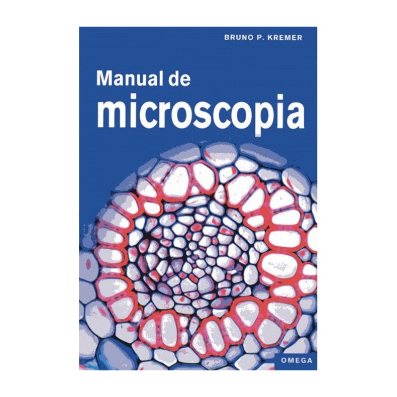
Author:Bruno P. Kremer
Publisher: PLANETA
Year: 2012
TABLE OF CONTENTS.
Introduction. Invitation to go beyond limits.
Journey to small worlds.Expeditions into the invisible.Shifting the boundaries of experience. Orders of magnitude in biology. A captivating new world.History of a look.At first it was the glasses. From the telescope to the microscope. Venetian glass and British lenses.
Exploration of inorganic nature.The simplest preparations.Trenches in the glass. A tremor: Brownian motion.Air bubbles in the preparation.Curved to all sides. Fully reflected. Diffraction waves.Microscopy of tiny crystals.Crystals from concentrated solutions. Crystallization by melting. Microsublimation: crystals reach the coating.Thin sections of hard materials.Rock samples under the microscope. In very thin sections. Traces of former life.Plastics and synthetic fibers.Reticulated cross for orientation in space. Nylon or perlon? Artificial silk or natural silk?Snow and frost crystals.Fixing flakes in lacquer. Direct snow. Frost: ice on the stem.
On the threshold of life.Phytoviruses.Tobacco mosaic virus. Viruses are not living beings. Parasites with the minimum size.Bacteria: the smallest living beings.Dental and lingual calculus. Protocytes are the simplest cells. Lactic acid bacteria.Cyanobacteria.Cells change the world. Loose and tight associations. Heterocysts are different.
The cell and its components.Basic cell plan.All living beings are cellular beings. Animal cells: simple but also complex. A microscopy classic: the epidermis of onion scales.The plant cell wall.Clear models: cell walls of the stem pith. Intercellular spaces: gaps between cells. Sequence of joined layers.Plastids: dyed or colorless.The internal structure. Temporary storage for the products of photosynthesis. Conversion to chromoplasts.The vacuoles: store and water reservoir.Anthocyanins or betalains. Abundant deposition. Plasmolysis: vacuoles may shrink.Chromosomes and nuclear division.To grow by division. Cutting, dyeing, pressing. Live observation.Membranes and mitochondria.Cellular power plant. Staining of mitochondria. Nerve cell endomembrane system.
Unicellular organisms and other protists.Plankton and small animals and plants.Infusion of hay. To help oneself from abundance. Fishing in open waters.Algae: slightly different plants.Massive seasonal development. One cell: four basic types. Green algae: representatives of various kinds.Euglenophyceae.Euglenophyceae: spindle-shaped or spherical. Green protozoa. Protists: neither plants nor animals.Colony formation in algae.A green algae with special mesh. Cellular chains and sprockets. Floating plates, rolling balls.Green algae on bark and rocks.Simple preparation. Species-rich growth. Aerial plankton algae.Diatoms.Cellular wall showcase. Construction as a cheese box. The lower part becomes a lid.Zignemataceae and Desmidiaceae.Twisted ribbons. Lamellar chloroplasts. Desmidiaceae: plastids with prominent figures.Seaweed.Stored liquid. Conservation by desiccation. Change of generations.Algae in symbiosis.Green animals. Stable union. Association with algae.Protozoa.Conceptual approach. The paradigm: the paramecia. Field of cilia.The diversity of protists.Foraminifera: with many pores. Changing forms. Amoebae with shells.
The kingdom of fungi.Uni- or multicellular microfungi.There is no beer without fungi. Yeasts are (almost) everywhere. Symbiotic yeasts on sucking insects.Filamentous microfungi.Molds: fungi in food. Mycelia in the shop window. Hyphae on adhesive tape: copy or protagonist.Fruiting bodies of macrofungi.Hats, caps, berets. Spores on the asci. Basidiomycetes with basidia.Fungi that produce mildew, blight and rust.Putrefaction. White coatings. Blackness: clandestine work of fungi.Lichens: dual organisms.Unicellular subtenants. The lichen as a dual organism. Different components.
Plants everywhere.Mosses: the simplest terrestrial plants.Mosses as a plant model. Placing leaves in flat position. Two types of leaves.Hepatic thallous.Segmented like a liver. Thallus multilayered. Gametangia umbrella-shaped.Sphagnos.Sphagnum in the pot. Green or colorless. Pores to the outside world.Ferns: archaic terrestrial plants.Sporulation: fecund without fruits. Reproductive change in two stages. Fertilization in water.Root anatomy.Longitudinal growth: maximum yield. Away from light: gravitropism and statolytic amyloplasts. Root vascular tissue: rays, laminae and cylinders.Vascular tissues in the stem.Vascular tissues in bundles. Water traveling upward. Pathways for sugars.Aerenchyma: plant gas pipelines.Stem pith inconsistent. Underdeveloped vascular bundles. Oxygen supply to the base.Log: cells, material, database.Libber and wood: soft bark, hard core. A look into the past. Medullary rays link inner and outer.Variety of leaves.Leaves always have several layers. The cuticle: hermetic seal. The epidermis as a showcase.Epidermis: facade of terrestrial plants.Isolated epidermis. Stomata design. Calcar.The needles are also leaves.Thin, but complex. Epidermal exoskeleton. Folded palisade and resiniferous ducts.The striking color of the flowers.Flowers as food suppliers: outdoor beverage kiosks. Target principle. Additive and subtractive color mixing.Pollen and pollen analysis.Collecting pollens. Fine preparations such as dust. Flying gametophytes.Fruits and seeds.Caryopsis going to the point. Pericarp and testa. Embryo: plant in compact form.
Of lower and higher animals.Elements of the skeleton of simple invertebrates.Glass skeletons. Leather and calcified. Spines in the skin.Arthropods: segmented animals.Insects: notched animals. Wings: colorful and cheerful. A look at the mouths of insects.Scales, shields, feathers, hairs.Surface view. Separation by layers. Pigmentation of the skin: up to blackening.Blood cells and blood groups.How to extract blood. Blue blood for technical reasons. White blood cells.The striated musculature.Muscle fibers to the extreme. The cable as a construction principle. Transverse and longitudinal grooves.
Methodology and techniques.Procurement of chemicals. Quantities. Concentrations. Time measurements. Safety aspects.Technical bases of work and preparation.The use of the microscope. Fresh or permanent preparation. Degreasing of slides.The inclusion of objects.Glycerin gelatin according to Kaiser (category A). Polyvinyl-lactophenol (category A). Polyhistol (category A).Staining and detection methods.Smear with India ink according to Burri. Observation of bacterial flagella. Staining of bacterial flagella according to Leifson.Methods of observation and illumination.Köhler and critical illumination. Brightfield illumination with transmitted light. Darkfield illumination with transmitted light.Cultivation methods.Cultivation collections. Soil decoction for algae culture. Standard culture media for microalgae.Immersion.
Bibliography. Useful addresses. Alphabetical index.
BOOK DETAILS
- Author(s): Bruno P. Kremer
- Binding: Paperback
- Printing: Color
- Year of issue: 2012
- Language of the book: Spanish
- Number of edition: 1
- Number of pages: 320
- Number of volumes: 1
- Translator/s: Joan Farré
- ISBN: 978-84-282-1570-1
- Measurements: 17 (width) x 25 (height)
